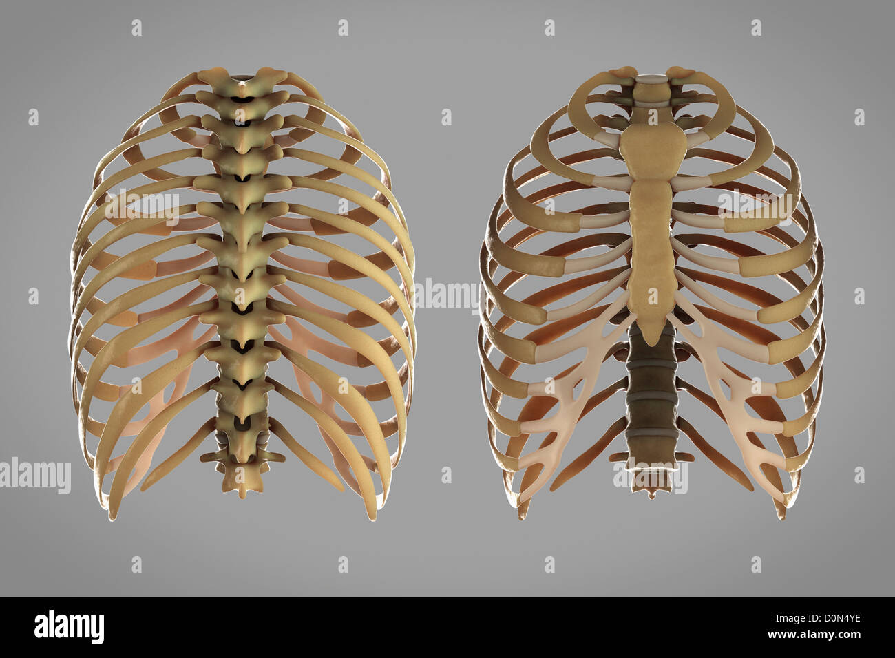
Anatomy Rib Cage Posterior View - Human Skeleton System Rib Cage Posterior View Anatomy Stock Illustration Illustration Of Anatomical Health 104471711 - An inhalation is accomplished when the muscular diaphragm, at the floor of the thoracic cavity, contracts and flattens, while the contraction of intercostal muscles lift the rib cage up and out.
byAdmin•
0
Anatomy Rib Cage Posterior View - Human Skeleton System Rib Cage Posterior View Anatomy Stock Illustration Illustration Of Anatomical Health 104471711 - An inhalation is accomplished when the muscular diaphragm, at the floor of the thoracic cavity, contracts and flattens, while the contraction of intercostal muscles lift the rib cage up and out.. It is formed by the 12 thoracic vertebrae, 12 pairs of ribs and associated costal cartilages and the sternum. This is because an ap view will exaggerate the heart size due to magnification. Gross anatomy (also called topographical anatomy, regional anatomy, or anthropotomy) is the study of anatomical structures that can be seen by unaided vision. Visceral pleura crosses mal at the level of the 8th rib). Lumbar (or 13th) ribs are a rare anatomical variant and represent transitional vertebrae at the thoracolumbar junction with a prevalence of ~1% 1.
Jul 27, 2021 · the thoracic cage (rib cage) is the skeleton of the thoracic wall. The rib cage also anchors the bones of the head, neck, shoulders, and arms to the trunk of the body. It presents as an additional rib coming off t13 or l1 (depending on numbering classification) and m. It is formed by the 12 thoracic vertebrae, 12 pairs of ribs and associated costal cartilages and the sternum. Powerful muscles that move the head and arms attach to these bones as well.
Thoracic Cage High Resolution Stock Photography And Images Alamy from c8.alamy.com Jul 27, 2021 · the thoracic cage (rib cage) is the skeleton of the thoracic wall. An inhalation is accomplished when the muscular diaphragm, at the floor of the thoracic cavity, contracts and flattens, while the contraction of intercostal muscles lift the rib cage up and out. Visceral pleura crosses mal at the level of the 8th rib). It presents as an additional rib coming off t13 or l1 (depending on numbering classification) and m. Both right and left pleural lines pass midaxillary line (mal) 12 th rib: 21.1 anatomy of the lymphatic and immune systems ; The visceral pleura remains roughly two ribs higher than the lines of pleural reflection in the lower thorax (e.g. Gross anatomy (also called topographical anatomy, regional anatomy, or anthropotomy) is the study of anatomical structures that can be seen by unaided vision.
The human rib cage is a component of the human respiratory system.
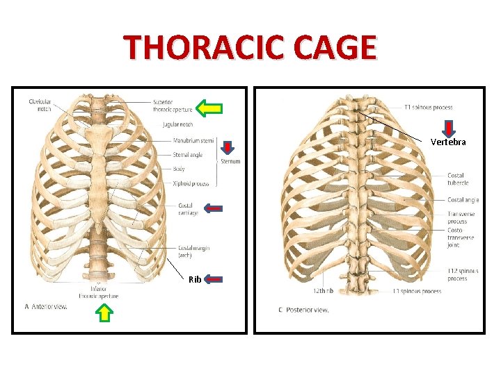
Both right and left pleural lines pass midaxillary line (mal) 12 th rib: It encloses the thoracic cavity, which contains the lungs. Lumbar (or 13th) ribs are a rare anatomical variant and represent transitional vertebrae at the thoracolumbar junction with a prevalence of ~1% 1. Gross anatomy (also called topographical anatomy, regional anatomy, or anthropotomy) is the study of anatomical structures that can be seen by unaided vision. Visceral pleura crosses mal at the level of the 8th rib). The regions of the body are labeled in boldface. It presents as an additional rib coming off t13 or l1 (depending on numbering classification) and m. Powerful muscles that move the head and arms attach to these bones as well. An inhalation is accomplished when the muscular diaphragm, at the floor of the thoracic cavity, contracts and flattens, while the contraction of intercostal muscles lift the rib cage up and out. However, a pa view is required to confidently diagnose cardiac enlargement. It is subdivided into gross anatomy and microscopic anatomy. The rib cage also anchors the bones of the head, neck, shoulders, and arms to the trunk of the body. Jul 03, 2018 · the bones of the chest and upper back combine to form the strong, protective rib cage around the vital thoracic organs such as the heart and lungs.
Powerful muscles that move the head and arms attach to these bones as well. Both right and left pleural lines pass midaxillary line (mal) 12 th rib: The human rib cage is a component of the human respiratory system. The visceral pleura remains roughly two ribs higher than the lines of pleural reflection in the lower thorax (e.g. This is because an ap view will exaggerate the heart size due to magnification.
Muscles Involved In Respiration Prof Ahmed Fathalla Ibrahim from slidetodoc.com The thoracic cage takes the form of a domed bird cage with the horizontal bars formed by ribs and costal cartilages. It encloses the thoracic cavity, which contains the lungs. The visceral pleura remains roughly two ribs higher than the lines of pleural reflection in the lower thorax (e.g. The human rib cage is a component of the human respiratory system. The rib cage also anchors the bones of the head, neck, shoulders, and arms to the trunk of the body. It is subdivided into gross anatomy and microscopic anatomy. Jul 27, 2021 · the thoracic cage (rib cage) is the skeleton of the thoracic wall. The regions of the body are labeled in boldface.
21.1 anatomy of the lymphatic and immune systems ;
The rib cage also anchors the bones of the head, neck, shoulders, and arms to the trunk of the body. Powerful muscles that move the head and arms attach to these bones as well. The thoracic cage takes the form of a domed bird cage with the horizontal bars formed by ribs and costal cartilages. Jul 03, 2018 · the bones of the chest and upper back combine to form the strong, protective rib cage around the vital thoracic organs such as the heart and lungs. Both right and left pleural lines travel posteriorly around the chest wall. An inhalation is accomplished when the muscular diaphragm, at the floor of the thoracic cavity, contracts and flattens, while the contraction of intercostal muscles lift the rib cage up and out. Gross anatomy (also called topographical anatomy, regional anatomy, or anthropotomy) is the study of anatomical structures that can be seen by unaided vision. Visceral pleura crosses mal at the level of the 8th rib). Lumbar (or 13th) ribs are a rare anatomical variant and represent transitional vertebrae at the thoracolumbar junction with a prevalence of ~1% 1. It is formed by the 12 thoracic vertebrae, 12 pairs of ribs and associated costal cartilages and the sternum. The human rib cage is a component of the human respiratory system. And it is enclosed by the rib cage. The regions of the body are labeled in boldface.
The human rib cage is a component of the human respiratory system. The rib cage also anchors the bones of the head, neck, shoulders, and arms to the trunk of the body. This is because an ap view will exaggerate the heart size due to magnification. Both right and left pleural lines pass midaxillary line (mal) 12 th rib: Jul 27, 2021 · the thoracic cage (rib cage) is the skeleton of the thoracic wall.
Muscles Involved In Respiration Prof Ahmed Fathalla Ibrahim from slidetodoc.com It is subdivided into gross anatomy and microscopic anatomy. The regions of the body are labeled in boldface. Lumbar (or 13th) ribs are a rare anatomical variant and represent transitional vertebrae at the thoracolumbar junction with a prevalence of ~1% 1. Jul 03, 2018 · the bones of the chest and upper back combine to form the strong, protective rib cage around the vital thoracic organs such as the heart and lungs. The visceral pleura remains roughly two ribs higher than the lines of pleural reflection in the lower thorax (e.g. An inhalation is accomplished when the muscular diaphragm, at the floor of the thoracic cavity, contracts and flattens, while the contraction of intercostal muscles lift the rib cage up and out. And it is enclosed by the rib cage. Visceral pleura crosses mal at the level of the 8th rib).
It is subdivided into gross anatomy and microscopic anatomy.
And it is enclosed by the rib cage. This is because an ap view will exaggerate the heart size due to magnification. Powerful muscles that move the head and arms attach to these bones as well. Jul 27, 2021 · the thoracic cage (rib cage) is the skeleton of the thoracic wall. An inhalation is accomplished when the muscular diaphragm, at the floor of the thoracic cavity, contracts and flattens, while the contraction of intercostal muscles lift the rib cage up and out. It is formed by the 12 thoracic vertebrae, 12 pairs of ribs and associated costal cartilages and the sternum. Gross anatomy (also called topographical anatomy, regional anatomy, or anthropotomy) is the study of anatomical structures that can be seen by unaided vision. It encloses the thoracic cavity, which contains the lungs. It is subdivided into gross anatomy and microscopic anatomy. 21.1 anatomy of the lymphatic and immune systems ; Visceral pleura crosses mal at the level of the 8th rib). Both right and left pleural lines travel posteriorly around the chest wall. The rib cage also anchors the bones of the head, neck, shoulders, and arms to the trunk of the body.
211 anatomy of the lymphatic and immune systems ; anatomy rib cage. Both right and left pleural lines travel posteriorly around the chest wall.